Article
Juvenile Chronic Arthritis
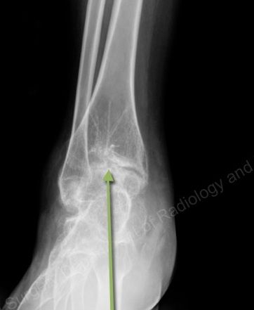
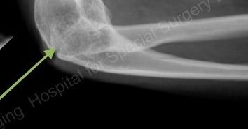
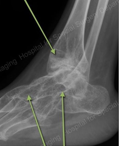
In a 30-year-old woman with decreased range of motion in several joints, radiographs show bony fusion, profound osteopenia, erosions and advanced arthrosis.
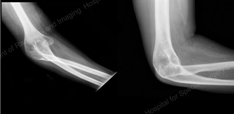
This imaging case study from the Hospital for Special Surgery assess a 30-year-old female with a history of decreased range of motion in multiple joints. The diagnosis is juvenile chronic arthritis.
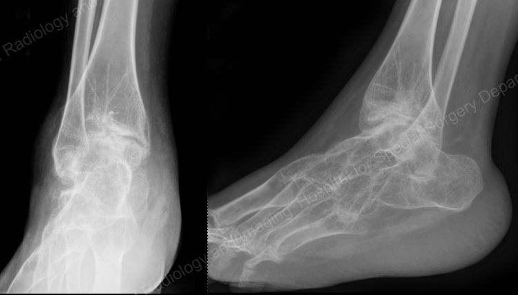
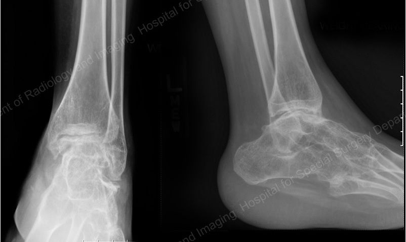
The images show AP and lateral radiographs of the right elbow (at left), right ankle (below, left), and left ankle (below, right).
FINDINGS
Radiographs of the elbow and both ankles show:
• a profound degree of osteopenia,
• bony fusion across multiple joints (arrows, below),
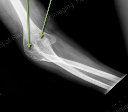
CLICK TO ENLARGE
Bony fusion of the radiocapitellar and humeroulnar joints.
• somewhat ballooned architecture to the ends of the bones (arrow, below),
CLICK TO ENLARGE
• long-standing radiocapitellar dislocation (arrow, below),
CLICK TO ENLARGE
Dislocation of the radiocapitellar joint
• and erosions and advanced arthrosis of both tibiotalar joints (below).
CLICK TO ENLARGE
DIAGNOSIS: Juvenile Chronic Arthritis (JCA)
JCA is a heterogeneous collection of inflammatory arthropathies occuring in patients less than 16 years of age. It is typically divided into juvenile onset ankylosing spondylitis, juvenile onset psoriasis/inflammatory bowel disease, juvenile onset adult type rheumatoid arhtirits (RA), and Still’s disease. Still’s disease is then separated into pauciarticular, polyarticular, or systemic variants. Systemic implies serositis, renal disease, hepatosplenomegaly, and lymphadenopathy.
The radiologic findings of JCA are similar to adult onset RA except that typically JCA will involve the larger joints first (knees, ankles, and elbow) and the smaller joints of the hand and wrist later.
The early findings of JCA are pronounced osteopenia and periarticular soft tissue swelling. With continued hyperemia to the joint there is a ballooning of the ends of the bones, and as the disease progresses, erosions and then ankylosis become present.
RESOURCES
• Resnick. Diagnosis of Bone and Joint Disorders. 4th Ed. 2002
• Brower. Arthritis in Black and White. 2nd Ed. 1997
• www.arthritis.co.za/jra.htm


