Article
Biomarkers Identify Patients at Risk for Hip Replacement Failure
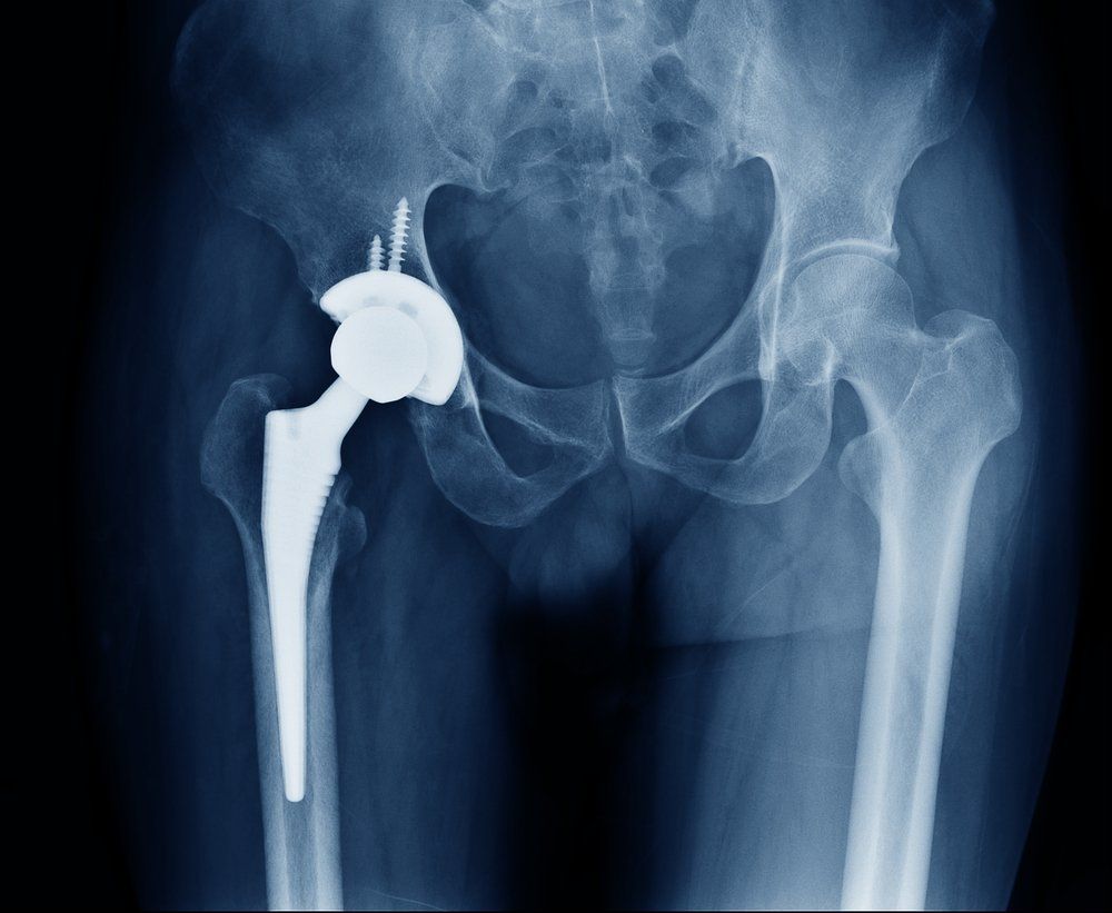
Urinary markers may predict the development of osteolysis following total hip replacement.
Image credit: ©ChooChin/Shutterstock.com

Urinary markers may predict the development of osteolysis in patients who have undergone total hip replacement, according to researchers at Rush University Medical Center in Chicago. Their findings are published in the Journal of Orthopaedic Research.
For the study, researchers used a repository of 24-hour urine samples collected before surgery and annually thereafter in 26 patients. Osteolysis eventually developed in 16 of these patients.
The levels of certain markers helped the investigators identify patients at risk for osteolysis long before the emergence of radiographic signs-in some cases, 6 years before a diagnosis was made. Although single markers showed moderate accuracy, the combination of α-crosslaps (α-CTX), a bone resorption marker, and interleukinâ6 (ILâ6), an inflammatory marker, led to high accuracy in the differentiation of patients who eventually developed osteolysis from those with no signs of osteolysis.
“We are hopeful that early biomarkers for implant loosening will alert surgeons to be especially vigilant in their follow-up of at-risk patients and may eventually lead to treatments delaying or avoiding the need for revision surgery,” said senior author Dr. D. Rick Sumner, of Rush University Medical Center. “Perhaps even more intriguing is that the two biomarkers we identified also differed before surgery among patients who eventually developed peri-implant osteolysis and those who did not, supporting the concept that other researchers have proposed of genetic risk factors for loosening.”
References:
Ross RD, Deng Y, Fang R, et al. Discovery of biomarkers to identify peri-implant osteolysis before radiographic diagnosis. J Orthop Res. 2018 Jun 5. doi: 10.1002/jor.24044. [Epub ahead of print]
Urinary markers predict bone problems after hip replacement [press release]. Hoboken, NJ: John Wiley & Sons, Inc.; June 6, 2018.


