Article
Breakthrough First Step in Brain-Based Fibromyalgia Diagnosis
Author(s):
Fibromyalgia is notoriously difficult to diagnose. But a new study from the University of Colorado Boulder (CU Boulder) may have found a brain signature that red flags the disease.

Fibromyalgia is notoriously difficult to diagnose. There is no specific blood test or biomarker that physicians can go off of, but instead the condition is characterized by chronic widespread musculoskeletal pain, along with fatigue and mood disorders. However, some healthcare providers still don’t think that fibromyalgia is a “real” condition. But a new study from the University of Colorado Boulder (CU Boulder) may have found a brain signature that red flags the disease.
“The novelty of this study is that it provides potential neuroimaging-based tools that can be used with new patients to inform about the degree of certain neural pathology underlying their pain symptoms,” lead author Marina López-Solà, PhD, a post-doctoral researcher in CU Boulder’s Cognitive and Affective Control Laboratory, said in a news release.
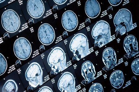
A total of 37 people with fibromyalgia and 35 healthy controls were exposed to painful pressure and non-painful visual, auditory, and tactile stimuli. The team used functional magnetic resonance imaging (fMRI) scans to evaluate brain activity during these scenarios.
“Though many pain specialists have established clinical procedures for diagnosing fibromyalgia, the clinical label does not explain what is happening neurologically and it does not reflect the full individuality of patients’ suffering,” said Tor Wager, PhD, director of the Cognitive and Affective Control Laboratory.
When the people with fibromyalgia were exposed to the painful stimuli, they had greater Neurologic Pain Signature (NPS) responses than those without the condition, as described in the journal PAIN. A new pain-related classifier, called FM-pain, emerged in sensory integration (insula/operculum) and self-referential (such as medical prefrontal) regions. In addition, there were reduced responses in the lateral frontal cortex in those with fibromyalgia.
The researchers also used a “multisensory” machine-learning techniques to identify brain-based fibromyalgia signs and non-painful sensory stimulation. When activity from NPS, FM-pain, and multisensory were combined, fibromyalgia was diagnosed with 92% sensitivity and 94% specificity.
While this may be a first step in such fibromyalgia research, it has the potential to be further developed in order to identify patient subtypes.
“The potential for brain measures like the ones we developed here is that they can tell us something about the particular brain abnormalities that drive an individual’s suffering. That can help us both recognize fibromyalgia for what it is — a disorder of the central nervous system – and treat it more effectively,” Wager concluded.
Related Coverage:
Disappointing Results in Adolescent Lyrica Study
EULAR’s Number One Recommendation for People with Fibromyalgia
Clinical Trial Confirms Swimming as Pain Soothing Exercise for Fibromyalgia


