Article
Image IQ: 7-year-old boy with photophobia
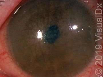
A mother brought her 7-year-old boy to the eye doctor after he complained for several months about increasing photophobia during outdoor activities like soccer practice and playground outings. She also noticed that he had been thirstier than usual during his soccer games, but wasn’t sweating as much as he usually did. During the slit lamp exam, the doctor noted crystals throughout his cornea and conjunctiva. What's your diagnosis?
(©VisualDx)

A mother brought her 7-year-old boy, who was a little small for his age, to the eye doctor after he complained for several months about increasing photophobia during outdoor activities like soccer practice and playground outings. He recently asked her to turn down the brightness level on his tablet when he was playing games. She also noticed that he had been thirstier than usual during his soccer games, but wasn’t sweating as much as he usually did. During the slit lamp exam, the doctor noted crystals throughout his cornea and conjunctiva.
What's your diagnosis?
A. Cystinosis
B. Conjunctivitis
C. Blepharospasm
D. Fungal corneal ulcer
Can you diagnose the patient? Use the differential builder on VisualDx: http://bit.ly/2ZHZFoW
See the next page for the answer.
The correct answer is A.) Cystinosis
Synopsis
Cystinosis is a clinically heterogeneous disorder with widespread organ damage resulting from tissue accumulation of cystine crystals. The most serious damage occurs in the kidney and may result in end-stage disease. However, other organs such as the thyroid and pancreas are often damaged as well.
Mutations in the CTNS gene are responsible for all cases of cystinosis, even though there can be considerable heterogeneity in clinical presentation. Individuals are generally grouped into 1 of 3 categories of disease. The most common and severe form is known as infantile or nephropathic cystinosis. These patients may have Fanconi-type renal tubular disease at an age as early as 6 months, and some patients require a renal transplant in the first decade of life. Poor feeding with failure to thrive are also evident in the first year of life. Retarded physical growth in the range of 50% of normal is evident in the same period. Hypophosphatemic rickets may be a consequence of untreated renal disease. Cystine crystals may be seen in the cornea at this time as well, and their presence causes significant sensitivity to light (photophobia). The retinal photoreceptors are damaged early, and patients may experience significant loss of vision as youngsters. Retinal pigment clumping and areas of hypopigmentation occur in the fundus periphery and progress centrally.
Intermediate or juvenile cystinosis is far less common, and the onset of clinical manifestations is often delayed until the second decade of life. Nephropathy is usually present but may be so mild that it can remain undetected until late stages. Electrolyte imbalance, growth retardation, and ocular symptoms may also escape detection for a decade or more. However, renal failure always occurs, usually by the third decade of life. These patients also have corneal crystals and photophobia and sometimes experience painful recurrent corneal erosions. Photoreceptor damage leading to vision loss is common. The pigmentary changes in the retina are similar to those in the infantile form of the disease.
Some individuals do not develop renal damage and have photophobia as their only symptom. This form of cystinosis is sometimes called ocular, non-nephropathic, or adult disease. Patients with this form of disease do not suffer photoreceptor damage or have retinal pigmentary changes. Corneal crystals cause photophobia, which may be the only symptom. Recurrent corneal erosions often occur.
Individuals with early-onset disease who survive into the third decade of life or later may develop late abnormalities such as myopathy, gastrointestinal dysfunction, cardiovascular disease, and central nervous system damage.
For more information about this diagnosis, visit VisualDx: http://bit.ly/2J6o6Wh

FDA Approves Crinecerfont for Congenital Adrenal Hyperplasia


