Article
Image IQ: Hemodialysis and a plaque on the leg
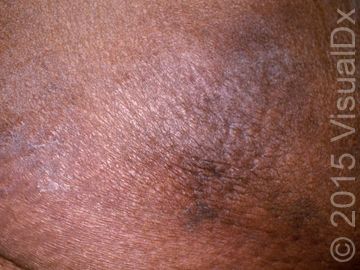
Image IQ: A 60-year-old woman undergoing hemodialysis for chronic kidney disease developes a smooth plaque on her leg. What's your diagnosis?
Image IQ: A 60-year-old woman undergoing hemodialysis for chronic kidney disease developes a smooth plaque on her leg. What's your diagnosis? (©VisualDx)

A 60-year-old woman currently undergoing hemodialysis for chronic kidney disease went to her physician when she saw a worrisome smooth plaque on her leg that developed over the last few weeks. It was very painful, especially when it first appeared. She thought that she had two more similar lesions developing and one looked sort of net-like.
What's your diagnosis? Use the differential builder on VisualDx to help select a diagnosis.
A. Cryoglobulinemia
B. Calciphylaxis
C. Cholesterol emboli
D. Antiphospholipid syndrome
See the next page for the answer.
ANSWER: B.) Calciphylaxis
Synopsis
Calciphylaxis (also called calcific uremic arteriolopathy) is a microvascular occlusion syndrome thought to be due to diffuse deposition of insoluble calcium salts in cutaneous blood vessels with associated thrombosis. While the exact pathogenesis is unclear, characteristic pathologic findings include progressive medial calcification of cutaneous blood vessels and subsequent ischemic necrosis of the skin. The process may be triggered by chronic hypocalcemia from decreased intestinal absorption of calcium, leading to increased levels of parathyroid hormone (PTH) and subsequent recruitment of calcium and phosphate from bone. Hypercoagulable states are also thought to play a possible role.
Calciphylaxis is increasing in incidence (5.7 per 10 000 chronic hemodialysis patients in 2011), and is most commonly associated with chronic renal failure, hemodialysis, and secondary hyperparathyroidism. There are also many cases of "nonuremic" or "nontraditional" calciphylaxis, which can occur in the setting of liver disease, diabetes, warfarin use, use of calcium-based phosphate binders, systemic corticosteroid use, solid organ malignancies, systemic lupus erythematosus, and Crohn disease. Other risk factors include female sex, obesity, Northern European descent, and hypoalbuminemia.
Notably, warfarin-associated nonuremic calciphylaxis tends to occur about 2.5 years after warfarin initiation on the lower extremities, does not have associated calcium abnormalities, and appears to have a more favorable prognosis than calciphylaxis associated with renal failure states.
Early lesions are extremely painful, violaceous retiform patches and plaques, classically on fat-bearing areas such as the thighs, buttocks, or abdomen. This is followed by necrosis, ulcers, eschar formation, and possibly gangrene. Mortality from calciphylaxis is high (60%-87%) and is largely secondary to sepsis from large, nonhealing ulcers.
For more information about this diagnosis and ICD 10 codes, visit Visual Dx.
DISCLOSURES: Image IQ courtesy of VisualDx.

FDA Approves Crinecerfont for Congenital Adrenal Hyperplasia


