Article
Image IQ: Smooth plaques with pain and itch
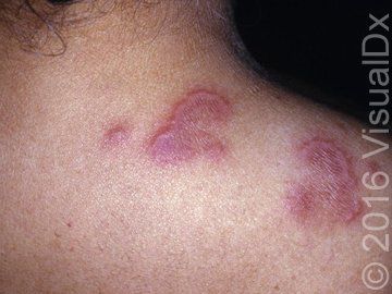
A 56-year-old woman with a history of celiac disease visited her doctor after a number of firm, smooth plaques developed over the course of a couple of weeks. The plaques manifested as yellow and pink, but then changed to a reddish color. What's the diagnosis?
(Image courtesy of VisualDx)

A 56-year-old woman with a history of celiac disease visited her doctor after a number of firm, smooth plaques developed over the course of a couple of weeks. The plaques manifested as yellow and pink, but then changed to a reddish color. They became painful and pruritic. The patient noted that the lesions seemed smaller in the morning and increased in size during the day. She was also experiencing joint pain.
Can you diagnose the patient? Use the differential builder in VisualDx as a guide.
A. Sweet syndrome
B. Erythema elevatum diutinum
C. Erythema multiforme
D. Ulcerative colitis
See the next page for the correct diagnosis.
The correct diagnosis is: B. Erythema elevatum diutinum
Erythema elevatum diutinum (EED) is a rare form of leukocytoclastic vasculitis in which immune complexes are deposited in small blood vessels, leading to inflammation. The exact etiology is unknown, but inciting factors may include infectious agents (streptococci, human immunodeficiency virus [HIV]), hematologic malignancies (monoclonal gammopathies), inflammatory bowel disease, celiac sprue, and rheumatologic disorders. EED may also be seen in association with immunoglobulin A (IgA) paraproteinemia. Case reports document associations with interferon-b, antituberculosis drugs, cisplatin, and erythropoietin.
Erythema elevatum diutinum presents as violaceous, erythematous, or brown papules, nodules, and/or plaques, predominantly over the extensor surfaces of the body. The most common associated symptom is arthralgia, although the lesions themselves may be painful or pruritic. Sometimes ocular abnormalities such as peripheral keratitis, panuveitis, and nodular scleritis have been seen. There is no racial predilection, but there is a slight female preponderance of disease. Incidence is highest in middle-aged to older adults, but may present earlier in those with HIV infection.
For ICD10 codes and to learn more about this diagnosis, visit VisualDx.


