Article
Knee pain in an 81-year-old man
An 81-year-old man with had a one-year history of right knee pain that had worsened in recent weeks despite no trauma or injury. What does the X-ray show?
An 81-year-old man presented to an urgent care facility with a 1-year history of pain in his right knee. He stated that the pain had become more severe over the previous 2 months but denied any acute trauma or injury. The physical examination revealed that the knee had limited range of motion.
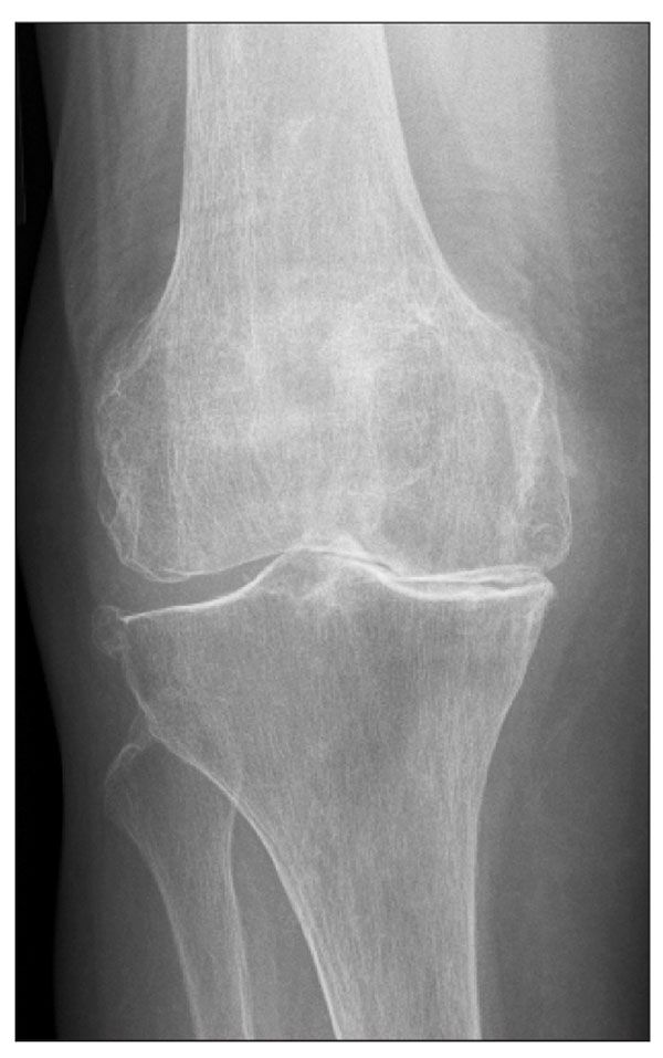
The accompanying anteroposterior x-ray view of the patient's right knee was obtained. What does it show? What is your interpretation?
The radiograph shows severe degenerative changes with joint-space narrowing most marked in the

medial compartment accompanied by subchondral sclerosis and osteophyte formation. No acute fracture is seen. The diagnosis was degenerative joint disease (DJD).
DJD significantly affects about 40 million persons in the United States.1 It is a progressive condition with "wear and tear of the articular cartilage"2 that becomes more severe with increasing age. The process involves joint-space narrowing, osteophyte formation, and sclerosis of the articular surface. The joints most often affected by DJD are the knees, first carpometacarpal joints, and hips.
Treatment for patients with DJD includes oral pain medications, anti-inflammatories, and surgical intervention. Conservative measures, such as weight loss, physical therapy with quadriceps strengthening, and realignment with bracing, also may help improve symptoms. DJD that affects the knees also may be managed with injection therapy that uses a short-acting analgesic combined with a corticosteroid or a viscosupplement. This treatment modality is best reserved for patients who want to delay surgery or for whom surgery is not recommended.
The patient was not a surgical candidate because of his comorbidities, which included chronic obstructive pulmonary disease and congestive heart failure. He was injected with a 21-gauge needle and a 5-mL syringe with 4 mL of 0.5% bupivacaine and 1 mL of methylprednisolone acetate, 40 mg/mL. The patient was advised to follow up for recheck after 1 month.
References:
1. Namba RS, Skinner HB, Gupta R. Adult reconstructive surgery. In: Skinner HB, ed. Current Diagnosis & Treatment in Orthopedics. 4th ed. New York: Lange Medical Books/McGraw-Hill. 2006:381-391.
2. Burnett S. Druginfonet.com. Degenerative Joint Disease. http://www.healthscout.com/ency/416/577/main.html. Updated July 11, 2005. Accessed September 22, 2009.


