Article
Nobel Prize in Chemistry Awarded for Cyro-Electrion Microscopy
Author(s):
Awarded to Jacques Dubochet, Joachim Frank, and Richard Henderson, the development has had a significant impact on medical research into infectious diseases.

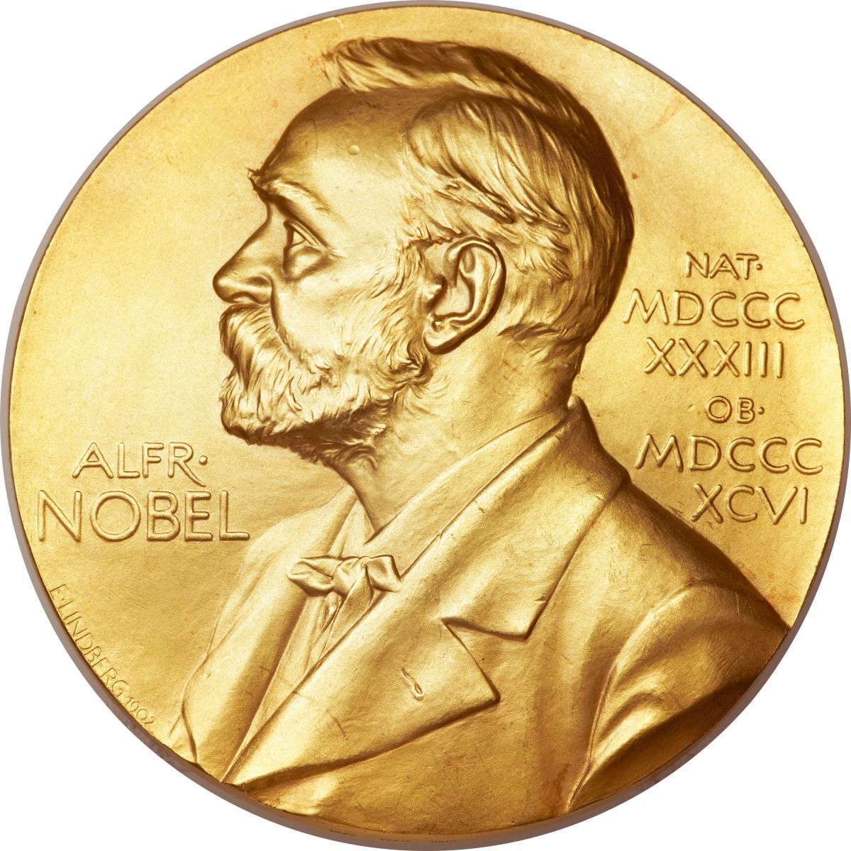
The Royal Swedish Academy of Sciences announced that it awarded the 2017 Nobel Prize in Chemistry to Jacques Dubochet, PhD, of the University of Lausanne; Joachim Frank, PhD, of Columbia University; and Richard Henderson, PhD,of the MRC Laboratory of Molecular Biology in Cambridge, for the development of cryo-electron microscopy for the high-resolution structure determination of biomolecules in solution.
As a result of the trio’s combined efforts, researchers can now regularly produce 3-D images and structures of biomolecules, which has had an enormous impact on the field of medicine and medical research.
The technology has allowed researchers to view proteins that result in antibiotic resistance, and have even been able to view the surfaces of viruses like Zika, resulting in a positive influence on research into infectious diseases.
The development has achieved both a simplification and improvement of the imaging in biomolecules. The method - which “moved biochemistry into a new era,” according to the Royal Swedish Academy of Sciences - works by freezing biomolecules mid-movement in order to visualize many processes that were otherwise unseen by science.
Henderson’s portion of the prize was awarded for his 1990 breakthrough in imaging with an electron microscope, using the device to produce a 3-dimensional image of a protein at atomic resolution. This was previously thought to be impossible, as the electron beam destroys the biological material.
Frank’s work between 1975 and 1986 proved that electron microscopes could be applied in general use. He developed an image processing method where a sharper, 3-dimensional image was rendered from the merging and analysis of blurrier, 2-dimensional images produced by the microscope.
The final step in developing the system begin with Dubochet’s success in vitrifying water in the 1980s, cooling it so quickly that it would solidify around a sample, thus allowing biomolecules to maintain their shape - even within a vacuum.
“Biochemistry is now facing an explosive development and is all set for an exciting future,” the Academy stated.


