Article
Pig Model Analysis Leads to Aortic Disease Pathology Discovery
Author(s):
Researchers have found distinctions between calcific aortic valve disease and atherosclerosis that could lead to improved care.

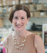
Kristyn Masters, PhD
An animal model-based study has uncovered explanation as to why common atherosclerosis therapies do not translate to treatment for calcific aortic valve disease (CAVD).
Researchers from the University of Wisconsin-Madison’s Arlington Agricultural Research Station have uncovered a new understanding of the early stages of stenosis — the severe narrowing of the aortic valve which results in reduced blood flow and a weakened heart. In analyzing these early stages, they have found key differences in the disease progressions for CAVD and atherosclerosis, and can now pursue therapies for the rare aortic disease that do not include statins or open-heart valve surgery.
The successful research is in part due to the use of pig organs as comparative human organ models for research that was previously impeded by a lack of candidate animal models. Because pigs, especially those bred with an overdose of artery fatty molecules, can better depict the early stages of human CAVD, researchers found that the accumulation of glycosaminoglycans (GAGs) in valve tissue indicates the beginning of CAVD.
Researchers then analyzed the tissue response to increased GAGs — a class of sugar molecules — through a novel platform that mimics the early pig CAVD process in a laboratory setting, with valve cells taken from their native healthy form. The result was a two-fold discovery: GAGs increased a chemical needed to grow new blood vessels, and it trapped low-density lipoprotein (LDL) molecules.
Because LDL molecules are trapped and eventually oxidized, their accumulation results in a multi-stage process that eventually causes valve cell damage in patients, researchers noted. The role of LDL molecules gives more understanding to the fact that 25% of adults aged 65 years or older have CAVD with partially blocked aortic valves, but only about 1% develop stenosis due to a valve being unable to open and close properly.
The discovery also indicates more distinction between CAVD and atherosclerosis in patients, as native valve cells are unable to oxidize LDL, while smooth muscle cells found in blood vessels can.
Prior to the research, it was commonly believed that CAVD was a valvular equivalent of atherosclerosis, Kristyn Masters, PhD, a professor of biomedical engineering at University of Wisconsin-Madison, said in a statement.
“Today, we know that valve cells are quite unique and distinct from the smooth muscle cells in our blood vessels, which explains why some treatments for atherosclerosis, such as statins, don’t work for CAVD, and why the search for drugs has to start from scratch,” Masters said.
The distinctions found in the study can serve as events to be targeted with new therapies for CAVD, Masters said. The goal is now to develop efficacious treatment that allows patients to valve replacement surgery. Part of that process is eased by this improved understanding of multiple steps, seen in a human-comparable, live model.
“The ability to examine multiple steps in this novel in vitro model for early CAVD opens up several promising avenues for developing drugs that are distinct from those for atherosclerosis,” Masters said.


