Article
Retinal Thinning May Be Linked to Vascular Alterations in AMD Patients
Author(s):
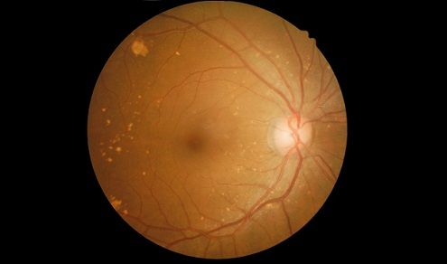
Recent research suggests that reticular pseudodrusen may be associated with the progression of early AMD to GA. Although an association has been theorized between pseudodrusen and AMD, researchers remain unclear on the pathogenesis of reticular pseudodrusen.

A Korean study has determined a possible link between retinal thinning and vascular alterations in early age-related macular degeneration (AMD) patients with reticular pseudodrusen.
The study, led by So Min Ahn, MD, with the Department of Ophthalmology at the Korea University College of Medicine in Seoul, retrospectively evaluated the clinical history and optical coherence tomography (OCT) and OCT angiography images of 135 early AMD eyes with reticular pseudodrusen. The goal of the study was to determine any relationship between retinal vessels and retinal thickness.
Recent research, Ahn told MD Magazine®, suggests that reticular pseudodrusen may be associated with the progression of early AMD to geographic atrophy (GA). Although an association has been theorized between pseudodrusen and AMD, researchers remain unclear on the pathogenesis of reticular pseudodrusen.
In order to determine a possible association, the researchers reviewed the medical records of 135 patients visiting the Korea University Medical Center Clinic between June 2016 and January 2018. The 135 patients, diagnosed with early- or intermediate-stage (according to Age-related Eye Disease Study [ARED] classifications) AMD, were divided into 2 groups according to appearance of reticular pseudodrusen (n= 60) or absence of reticular pseudodrusen (75) on OCT images.
Choroidal thickness, vascular density of capillary plexus (superficial and deep), and foveal avascular zone measurements were completed for each of the 135 patients, and demographic and medical histories were obtained for analysis.
Ahn and colleagues discovered that, although there were no statistically significant differences between the vascular density or retinal thickness between the 2 groups, there were some marked differences in subfovial choroidal thickness between the groups. Multivariate analysis of data showed some statistically significant associations between vascular thickness and retinal thinning in the reticular pseudodrusen group, and age and retinal thickness in the non-reticular pseudodrusen group.
Mean retinal thickness was comparable between the 2 groups with the reticular pseudodrusen group at a mean 287.31 ± 24.36 µm and the non-reticular pseudodrusen group at a mean 294.27 ± 20.71 µm (P = 0.075).
Subfovial choroidal thickness varied significantly between the 2 groups with the mean subfovial choroidal thickness of the reticular pseudodrusen group at 158.13 ± 42.53 µm versus the non-reticular pseudodrusen group at 237.89 ± 60.94 µm (P < 0.001).
Researchers wrote that, in spite of the similarity of retinal thickness and vascular densities of superficial and deep capillary plexus, “different associations of retinal thickness and vascular density were observed between the 2 groups." The association suggests that retinal thinning in early AMD eyes may progress differently, and have different features, when reticular pseudodrusen are present.
In eyes without reticular pseudodrusen age itself may be contributing more to retinal thinning, which confirms earlier research suggesting that advancements in age contribute to the amount of choroidal atrophy in early AMD eyes.
Ahn suggested 2 possibilities arise from the analyses. The first is that eyes with reticular pseudodrusen under chronic choroidal insufficiency could contribute to retinal atrophy by decreasing metabolic demand and vascular flow.
The second, researchers wrote, is that "eyes with reticular pseudodrusen may be co-morbid for choroidal insufficiency and retinal vascular dysregulation" in which the blood flow to the retina and choroid are out of sync, increasing ocular perfusion pressure and inconsistent blood flow.
Ahn noted that further study is needed to determine the exact relationship of the association between retinal thinning and vascular alterations, but that the study data may suggest that retinal atrophy in eyes with reticular pseudodrusen may be affected by a blood flow impairment connected to the pathology of macular diseases like AMD.
The study, "Retinal vascular flow and choroidal thickness in eyes with early age-related macular degeneration with reticular pseudodrusen," was published online in BMC Ophthalmology last month.


