Article
Shoulder Pain in Older Patients: Interpreting Clinical Findings
Older patients experience shoulder pain more frequently than younger patients and present with different issues. Using a combination of tests with a detailed history is the best approach to narrowing the differential.
Older patients experience shoulder pain more frequently than younger patients, they present with different issues and concerns, and their treatment involves different considerations. As a result, physicians face special challenges in making the diagnosis and providing appropriate care for this patient population.
Once a thorough, relevant history is obtained and the physical examination is completed, knowing how to interpret clinical findings is important. Single physical examination maneuvers for making the diagnosis of shoulder conditions are not sensitive enough to rule out a problem or specific enough to rule one in. Some tests help decrease the likelihood of a patient having certain conditions, but the physician needs to use all the information obtained from the history and physical examination to estimate the overall probability.1
This 2-part article describes our approach to identifying shoulder pain in older patients. In the first part (The Journal of Musculoskeletal Medicine, June 2009, page 216), we focused on the use of historical clues, a thorough physical examination, and testing to help narrow the differential diagnosis. This second part discusses interpretation of clinical findings.
Impingement syndrome, tendinopathy, and bursitis
Neer introduced the concept of the impingement syndrome in 1972. He found evidence during his dissections of mechanical impingement of the rotator cuff and humeral head against the anterior process of the acromion, along with traction of the coracoacromial ligament. He also described stages of impingement, with a continuum from edema and hemorrhage to fibrosis and tendinitis to bone spurs and tendon rupture.2
Neer and others considered the anteroinferior acromion, the coracoacromial ligament, and the acromioclavicular (AC) joint to play a role in impingement.3 However, other theories suggest that it is the rotator cuff that undergoes degeneration, leading to weakness and loss of forces and, subsequently, superior migration of the humeral head. The result is impingement, as well as subacromial changes. The cause of impingement probably is multifactorial-no single factor alone produces lesions or disorders of the rotator cuff.2-4
The subacromial bursa may become inflamed, resulting in shoulder pain; tendinopathy of the supraspinatus tendon also may be a source of pain. Active and passive motions of the shoulder often are limited because of pain in both conditions; the active component is much more limited than the passive one.5
Overlap of many clinical findings with impingement, tendinopathy, and bursitis is common. Findings of pain with active and, to some degree, passive motion also may be seen with impingement. Pain related to all 3 conditions usually occurs over the anterolateral aspect of the shoulder, often with some radiation to, but not usually beyond, the elbow.6
There also may be pain with manual muscle testing and complaints of pain at night. Pain at rest is more likely to be associated with bursitis and tendinopathy than with impingement, although such pain alone is not diagnostic.5
Chronicity and history are the most helpful components in differentiating bursitis, impingement, and tendinopathy. They have common treatment plans that focus on regaining normal and efficient shoulder movements, with emphasis on the scapula stabilizers and rotator cuff muscles.

On examination, a positive Neer sign, Hawkins sign, and painful arc are suggestive of impingement. Several studies have evaluated the sensitivity, specificity, and likelihood ratios (LRs) of these tests (Table 1).7,8 LRs combine sensitivity and specificity information; positive and negative LRs provide an indication of how much the odds of having a disease increase or decrease with a positive or negative test result, respectively.
The statistics indicate that these tests do not have good results consistent across the studies and a positive test result is not as helpful as a negative result. If the results are negative, the probability that the patient has impingement is low, but the positive tests are not as helpful individually.
As with all special testing at the shoulder, using a combination of tests with a detailed history is the best approach to narrowing the differential.7,8 The results of a combination of 7 tests for impingement (Hawkins sign, Neer sign, painful arc, drop arm sign, cross chest adduction test, Speed test, and Yergason test), if all positive, had a sensitivity and specificity of 4% and 97%, respectively; if at least 3 results were positive, the sensitivity and specificity were 84% and 44%, respectively.7 Because no one test alone has been shown to be reliably and reproducibly predictive of impingement syndrome, more studies are needed to better identify the best test combinations.
Rotator cuff tears
Tears may occur after a specific incident (eg, trauma or a fall) or with heavy lifting but also may develop over time in a more degenerative manner. The incidence of rotator cuff tears increases linearly with age-33%, 55%, 65%, and 80% in the 40s, 50s, 60s, and 70s, respectively.9
Yamaguchi and associates10 evaluated the contralateral, asymptomatic shoulder in patients who showed evidence of a unilateral rotator cuff tear on ultrasonography. If there was a full-thickness tear on the symptomatic side, there was a 35.5% chance of a full-thickness tear on the uninvolved side, a 20.8% chance of a partial-thickness tear, and a 43.7% chance of normal findings. If the painful side did not have a torn rotator cuff, there was little chance of a tear on the asymptomatic side (normal, 97.7%; full-thickness tear, 0.5%; partial-thickness tear, 1.8%). These findings should increase physicians’ awareness that specific age-groups are at higher risk than others. In addition, awareness of the prevalence of bilateral disease in those with unilateral rotator cuff tears may contribute to future evaluations and patient education.11
Rotator cuff tears are not always symptomatic. When pain is present, it is characteristically found at the lateral, anterior, and superior portions of the shoulder. Pain typically is not present at rest and is aggravated by abducting the arm.5
On examination, the patient often has a limitation or inability to actively raise the arm. Shrugging (“hiking”) the shoulder on attempting to raise the arm may be observed because the patient uses his or her upper trapezius muscles to accommodate for weakness in the supraspinatus. On inspection of the posterior portion of the shoulder and scapula, atrophy of the supraspinatus and infraspinatus might be seen, depending on the chronicity and severity of the tear. In older patients, 81% of those with atrophy had a rotator cuff tear; if atrophy is absent, however, this information is not useful, as evidenced by a negative predictive value of 43%.11

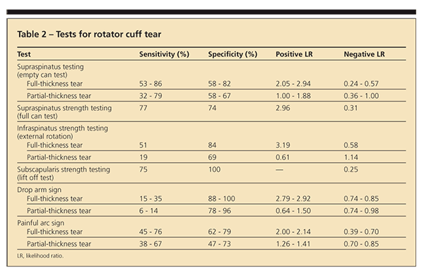
Evaluating for weakness on manual muscle testing may help identify a tear (Table 2), although multiple studies show a wide range of results.7-9,12,13 For supraspinatus and infraspinatus strength testing, particularly for full-thickness rotator cuff tears, negative results usually are helpful in that the likelihood of a tear is very low. Positive test results for the supraspinatus are inconsistent and less indicative. In the presence of a partial-thickness rotator cuff tear, the same tests are less helpful. For subscapularis tests, 1 study showed fair sensitivity and high specificity. Patients with rotator cuff tears also may have a positive Neer sign and positive Hawkins test result, a painful arc, and pain with active and passive motion.
Because of overlap of signs, symptoms, and provocative test results, using a combination of tests and clinical findings may help narrow the differential.9,11 Park and colleagues8 reported that if the
results of the painful arc test, drop arm sign, and infraspinatus strength test all are positive, the LR of a rotator cuff tear occurring is 15.57; if 2, 1, or none of the results is positive, the LR drops to 3.57, 0.79, or 0.16, respectively.
Adhesive capsulitis
Patients with adhesive capsulitis typically present with an insidious onset of a diffusely painful shoulder and stiffness. Sleep often is disrupted because of pain. Shoulder stiffness helps distinguish this cause from others. There often is significant restriction in active and passive motion in all directions (50% to 60%).5
If the patient’s active motion is limited during the examination, evaluating his passive range is important; limitation is suggestive of adhesive capsulitis. Patients may have pain with impingement testing and manual muscle testing, but range of motion is the key. Limitation of passive motion in any 1 range (external rotation, internal rotation, abduction, or forward elevation) is important to identify. The diagnosis is based primarily on the history and physical examination.
Three phases of adhesive capsulitis have been described: (1) patients typically have increasing stiffness and pain for 2 to 9 months, (2) they have substantial stiffness but less pain for 4 to 12 months, and (3) pain resolves and function gradually is restored for 5 to 26 months.5 Physical therapy is an important component of the treatment plan. The results of an Arslan and Celiker14 study comparing physical therapy and NSAIDs with an intra-articular corticosteroid injection for adhesive capsulitis showed no significant difference in outcomes. However, Carette and associates15 compared a single intra-articular glenohumeral corticosteroid injection, physical therapy alone, a combination of the 2, and placebo; the combination of physical therapy and intra-articular injection showed greater improvements in ranges of motion at 6 weeks and 3 months.
Evidence indicates that an intra-articular viscosupplemention injection may be helpful in some cases of adhesive capsulitis.16 Surgical intervention is necessary in patients for whom conservative management is not successful.
Adhesive capsulitis may be idiopathic or may result from underlying pathology, such as impingement or a rotator cuff tear. It also may present in patients who have an endocrinopathy, such as
diabetes mellitus or hypothyroidism, or as a result of trauma, including postsurgical trauma; these cases may require more aggressive management.
Osteoarthritis
Glenohumeral arthritis may be primary or secondary. For primary arthritis, the cause is unknown. Secondary causes include congenital or developmental abnormalities, infection, rheumatologic disorders (other than primary osteoarthritis), endocrine disorders, post-traumatic concerns, and neuropathic problems.17 There is a gradual, progressive mechanical and biochemical breakdown of articular cartilage and other tissues in the joint.18
Risk factors for osteoarthritis include age older than 50 years, crystals present in the joint, a history of injury to the joint, joint hypermobility or instability, and prolonged occupational or sports-related stress. Patients present with progressive activity-related pain, pain deep in the shoulder, and night pain; as the disease progresses, they experience pain at rest and worsening joint stiffness.
Early in the disease process, the pain and symptoms are mild and the examination results may be normal. As the disease becomes more advanced, both active and passive motions are lost, crepitus and grinding may be palpable on examination, and swelling and joint enlargement may be present.17,18 Radiographic findings for arthritis in the glenohumeral joint are similar to those for arthritis in other joints; they include joint-space narrowing, osteophyte formation, pseudocysts in the subchondral bone, and sclerosis.
Treatment varies with the degree of arthritis present and of functional limitation. The goals are to decrease pain, increase function, and improve the quality of life.
Acromioclavicular joint injury
Pain at the AC joint may result from a sprain or separation of the AC joint caused by disruption of the coracoclavicular ligament; both probably are post-traumatic. The presence of osteoarthritis or rheumatoid arthritis also could cause pain in this joint.2

On examination, there may be tenderness over the AC joint and localized swelling. Tenderness to palpation of the AC joint has been evaluated (Table 3).19,20 A positive result of the cross-body adduction test with pain localized over the AC joint suggests that the AC joint is the source of pain. If pain is reproduced over the AC joint with the O’Brien test, AC joint pathology could be indicated. There is low sensitivity and high specificity with this test, as well as wide variability in the LRs, making the results difficult to interpret.
Biceps tendinopathy
Tendinopathy of the long head of the biceps may be a source of shoulder pain in isolation or with other shoulder pathology (eg, impingement or a rotator cuff tear). Pain with biceps tendinopathy is located at the anterior aspect of the shoulder; it may be reproduced with palpation of the long head of the biceps tendon proximally along the bicipital groove.5
Pain with the Speed test is thought to be suggestive, but no studies have been found that look solely at this test and biceps tenosynovitis. Bennett21 study data for the Speed test with results for biceps and labral pathology show a sensitivity of 90%, specificity of 14%, positive LR of 1.05, and negative LR of 0.71. On the basis of the data and the lack thereof, the Speed test is difficult to interpret from the evidence with a positive or negative result.
Conclusions
Shoulder pain presents a diagnostic challenge because there are many possible causes. No one test is sensitive or specific enough to make a diagnosis. Special tests and maneuvers help when the results are negative and less so when they are positive. A combination of tests with several positive results may be most helpful.
References:
References1. Ebell M. Diagnosing rotator cuff tears. Am Fam Physician. 2005;71:1587-1588.
2. Steinfeld R, Valente RM, Stuart MJ. A commonsense approach to shoulder problems. Mayo Clin Proc. 1999;74:785-794.
3. Neviaser RJ, Neviaser TJ. Observations on impingement. Clin Orthop Relat Res. 1990;254:60-63.
4. Miller RH II, Dlabach JA. Shoulder and elbow injuries. In: Canale ST, Beaty JH, eds. Campbell’s Operative Orthopaedics. Vol 3. 11th ed. Philadelphia: Mosby; 2007:2609.
5. Andrews JR. Diagnosis and treatment of chronic painful shoulder: review of nonsurgical interventions. Arthroscopy. 2005;21:333-347.
6. Woodward TW, Best TM. The painful shoulder, part II: acute and chronic disorders. Am Fam Physician. 2000;61:3291-3300.
7. Calis M, Akgün K, Birtane M, et al. Diagnostic values of clinical diagnostic tests in subacromial impingement syndrome. Ann Rheum Dis. 2000;59:44-47.
8. Park HB, Yokota A, Gill HS, et al. Diagnostic accuracy of clinical tests for the different degrees of subacromial impingement syndrome. J Bone Joint Surg. 2005;87A:1446-1455.
9. Murrell GA, Walton JR. Diagnosis of rotator cuff tears. Lancet. 2001;357:769-770.
10. Yamaguchi K, Ditsios K, Middleton WD, et al. The demographic and morphological features of rotator cuff disease: a comparison of asymptomatic and symptomatic shoulders. J Bone Joint Surg. 2006;88A:1699-1704.
11. Litaker D, Pioro M, El Bilbeisi H, Brems J. Returning to the bedside: using the history and physical examination to identify rotator cuff tears. J Am Geriatr Soc. 2000;48:1633-1637.
12. Itoi E, Kido T, Sano A, et al. Which is more useful, the “full can test” or the “empty can test,” in detecting the torn supraspinatus tendon? Am J Sports Med. 1999;27:65-68.
13. Hertel R, Ballmer FT, Lombert SM, Gerber C. Lag signs in the diagnosis of rotator cuff rupture. J Shoulder Elbow Surg. 1996;5:307-313.
14. Arslan S, Celiker R. Comparison of the efficacy of local corticosteroid injection and physical therapy for the treatment of adhesive capsulitis. Rheumatol Int. 2001;21:20-23.
15. Carette S, Moffet H, Tardif J, et al. Intraarticular corticosteroids, supervised physiotherapy, or a combination of the two in the treatment of adhesive capsulitis of the shoulder: a placebo-controlled trial. Arthritis Rheum. 2003;48:829-838.
16. Calis M, Demir H, Ulker S, et al. Is intraarticular sodium hyaluronate injection an alternative treatment in patients with adhesive capsulitis? Rheumatol Int. 2006;26:536-540.
17. Hinton R, Moody RL, Davis AW, Thomas SF. Osteoarthritis: diagnosis and therapeutic considerations. Am Fam Physician. 2002;65:841-848.
18. Millett PJ, Gobezie R, Boykin RE. Shoulder osteoarthritis: diagnosis and management. Am Fam Physician. 2008;78:605-611.
19. Walton J, Mahajan S, Paxinos A, et al. Diagnostic values of tests for acromioclavicular joint pain. J Bone Joint Surg. 2004;86A:807-812.
20. Chronopoulos E, Kim TK, Park HB, et al. Diagnostic value of physical tests for isolated chronic acromioclavicular lesions. Am J Sports Med. 2004;32:655-661.
21. Bennett WF. Specificity of the Speed’s test: arthroscopic technique for evaluating the biceps tendon at the level of the bicipital groove. Arthroscopy. 1998;14:789-796.


