Article
Smartphone Eye Exam for Diabetic Retinopathy
Scientists at the University of Bonn and Sankara Eye Hospital Bangalore (India) have jointly developed an inexpensive method of screening for vascular changes in the eyes of diabetics- using a smartphone.
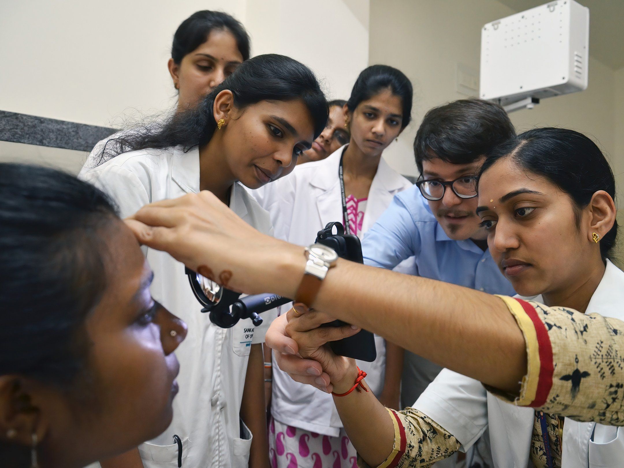
Dr. Maximilian W. M. Wintergerst (second from right) trains ophthalmic assistants at the Sankara Eye Hospital in Bangalore, India. PHOTO CREDIT© Universität Bonn/Sankara Eye Foundation

Scientists at the University of Bonn and Sankara Eye Hospital Bangalore (India) have jointly developed an inexpensive method of screening for vascular changes in the eyes of diabetics- using a smartphone.
Retinal damage due to diabetes is a common cause of blindness in working-age adults. It’s estimated that 80% of people with diabetes worldwide live in developing and emerging countries, and in many of these low- and middle-income countries, an eye examination in a physician’s office is not always possible. This makes systematic retinal screening of diabetics difficult in areas with few medical resources.
However, a new study published in the journal Ophthalmology, demonstrates that high resolution images from smartphones could help detect changes early.
In the study, the researchers compared four different approaches aimed at enabling ophthalmoscopy with a standard mid-range smartphone. "The best result in our test was achieved by an adapter with an additional lens that is attached to the smartphone," explained Dr. Maximilian Wintergerst from the Department of Ophthalmology at the University Hospital Bonn.
"It allowed almost 80 percent of eyes with any retinal changes to be detected, even in the early stages. Advanced damage could even be diagnosed 100 percent of the time."
Trained optometrists (ophthalmic assistants) from the Sankara Eye Hospital in Bangalore assisted in the study. On average, they needed one to two minutes per examination. This involved documenting changes in the retina by filming the back of the eye with a smartphone camera.
Study co-author, Prof. Dr. Robert Finger from the Department of Ophthalmology at the University Hospital Bonn, considers these capabilities to be what makes the method so appealing: "This means that the examination can also be enabled by trained laypersons," he says. "The images are then sent via the Internet to the ophthalmologist for diagnosis."
COVID-19 has created additional need to explore methods of reducing hospital visits – the authors point out, and this method could eventually be useful in reducing physician-patient contact.
The researchers are currently developing an app that will make it possible to create an encrypted electronic patient file for each patient on the smartphones used for the examination. The app will store the images as well the findings of the doctor who ultimately reviewed them. Future research will include the addition of automatic pre-evaluation of the images using artificial intelligence.
Publication: Maximilian W. M. Wintergerst, Divyansh K. Mishra, Laura Hartmann, Payal Shah, Vinaya K. Konana, Pradeep Sagar, Moritz Berger, Kaushik Murali, Frank G. Holz, Mahesh P. Shanmugam*, Robert P. Finger*: Diabetic retinopathy screening using smartphone-based fundus imaging in India; Ophthalmology, *these authors contributed equally, DOI: 10.1016/j.ophtha.2020.05.025 http://www.aaojournal.org/article/S0161-6420(20)30463-2/fulltext


