Article
45-Year-Old Woman With Longstanding Knee Pain
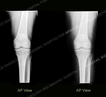
Images taken 7 months apart show subtle densities of the left knee that has been causing pain. The woman also has subluxation of the fingers, without erosion.

These images, taken 7 months apart, show the left knee that has been causing a 45-year-old woman to endure substantial pain for a very long time.
The one at left shows subtle densities that are more conspicuous on the later image, at right, with a serpiginous, well defined, sclerotic border.
Hand radiographs show ulnar subluxation of the metacarpophalaneal joints, without erosion.
What would you look for on MRI? What diagnosis would you predict?
Click here to read the full case study from the collection at New York's Hospital for Special Surgery.


