Article
Higher Prevalence of Diabetic Retinopathy Observed in Patients with T2D, Diabetic Nephropathy
Author(s):
Logistic regression analysis indicated that higher stages of urine albumin creatine ratio were significantly associated with diabetic retinopathy in patients with T2D and diabetic nephropathy.
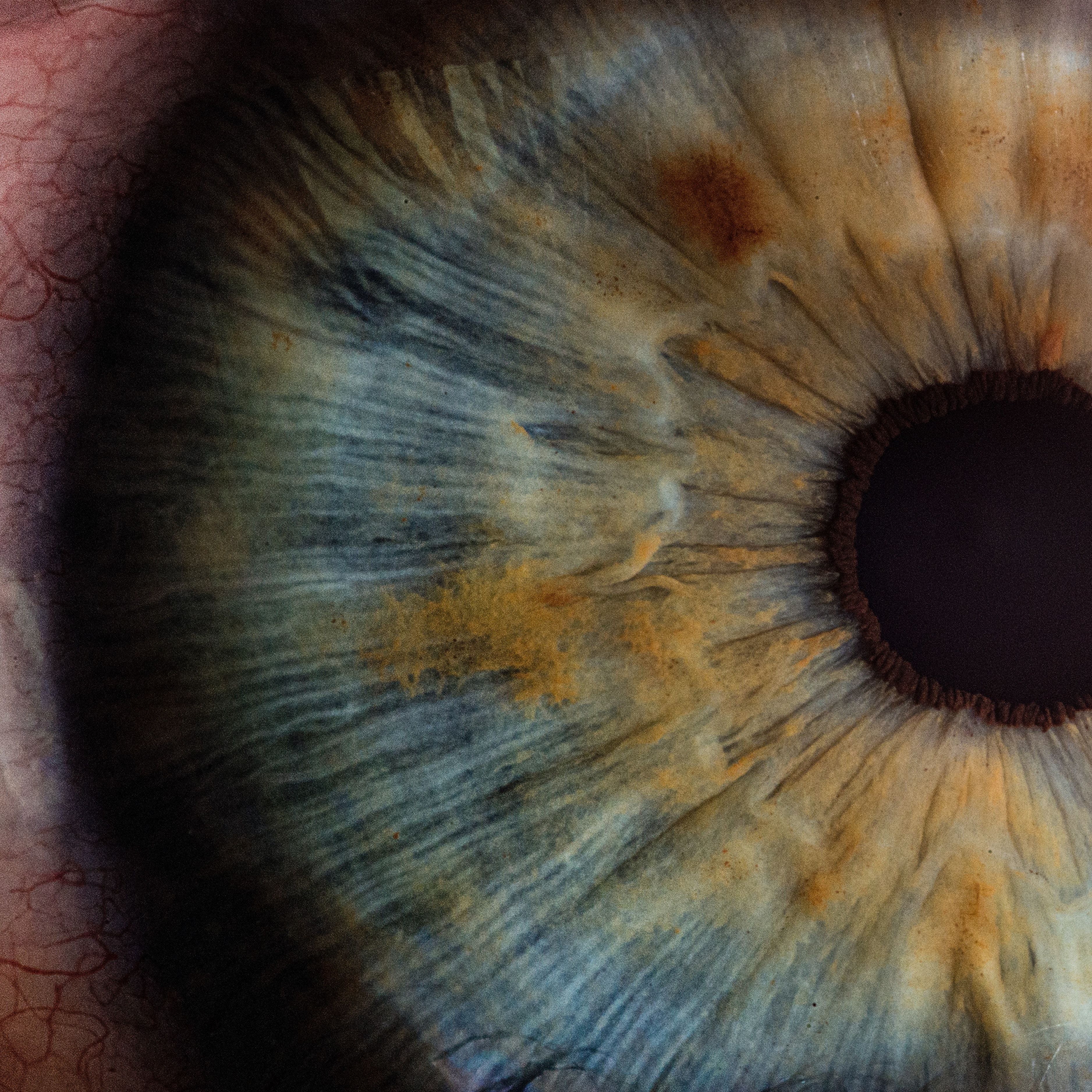
Credit: v2osk/Unsplash

New results from a retrospective, observational study found a relatively higher prevalence of diabetic retinopathy (DR) among patients with type 2 diabetes mellitus (T2D) with diabetic nephropathy.1
The study of nearly 150 patients with T2D suggested diabetes was significantly associated with the urine albumin creatine ratio (UACR) stage; thus, the investigative team from China-Japan Friendship Hospital suggested the ACR stage could be identified as a risk factor for DR in patients with diabetic nephropathy.
“Besides routine ophthalmic examination in patients with T2D, patients with diabetic nephropathy need ophthalmic examination more timely and more frequently,” investigators wrote.1 “These data may provide useful information for the prevention and management of diabetes complications.”
Recent literature has indicated that DR was significantly associated with renal function deterioration and those with diabetic nephropathy experienced a higher incidence of DR, compared with patients without DR.2 Noting that all diabetic microvascular complications share similar etiological factors, screening for DR should be taken in patients with diabetic nephropathy.
The investigative team, led by Hongsong Zhang, Department of Ophthalmology, China-Japan Friendship Hospital, and Zhijun Wang, Department of Endocrinology, China-Japan Friendship Hospital, hypothesized that the severity of diabetic nephropathy may be a potential risk factor for microvascular changes in the retina. They set out to test their hypothesis by exploring the characteristics of retina microvascular changes in patients with diabetic nephropathy and its risk factors.
The medical records of 145 patients diagnosed with T2D and diabetic nephropathy at China-Japan Friendship Hospital from January 2019 to December 2019 were retrospectively reviewed. Investigators acquired demographic and clinical parameters from medical records, including age, sex, duration of diabetes history, HbA1c level, and body mass index (BMI). The presence of DR was additionally examined and graded as absent DR, mild non-proliferative DR (NPDR), moderate NPDR, severe NPDR, and proliferative diabetic retinopathy (PDR).
All included patients had a thorough ophthalmic examination, including best-corrected visual acuity (BCVA) using a Snellen chart, intraocular pressure (IOP) measurement, slit lamp examination, color fundus photography, optical coherence tomography (OCT), and fluorescein angiography (FFA) if needed. The presence of hard exudates was made by visible white-yellow exudates in the posterior area in color fundus images.
A total of 140 eyes of 140 patients with T2D and a history of diabetic nephropathy were included in the analysis, with 86 (61.4%) eyes having DR. Meanwhile, PDR accounted for 23.6% (n = 33 eyes) in the 140 eyes and sight-threatening DR accounted for 35.7% (n = 50 eyes) of eyes. The analysis of laboratory profiles found no significant difference between DR group and non-DR group in triglyceride level and total and high-density lipoprotein cholesterol (HDL-C).
However, patients with DR had significantly higher levels of low-density lipoprotein cholesterol (LDL-C [P = .004]), HbA1c (P = .037), UACR (P <.0001), and lower levels of estimated glomerular filtration rate (eGFR [P = .013]). A logistic regression analysis indicated the presence of DR was significantly associated with UACR stage (P = .011). Those with UACR stage 3 were observed to have a higher incidence of DR, compared with patients with UACR stage 1 (OR, 24.15; 95% CI, 2.06 - 282.95).
Due to 7 patients lacking clear OCT images, 138 eyes of 138 patients with clear OCT images were analyzed for hard exudates and diabetic macular edema (DME). The analysis showed 32 eyes (23.2%) had hard exudates in the posterior pole and 13 eyes (9.4%) had DME. Between patients with hard exudates and those without hard exudates, investigators noted significant differences in the LDL-C level (P = .008), total cholesterol level (P = .014), and UACR (P = .001).
The analysis found that visual acuity was worse in the group with hard exudates than in the non-hard exudates group but without significant differences between groups (P = .058). However, there were significant differences observed in the LDL-C level and UACR between the DME group (P = .020) and the non-DME group (P = .009).
Investigators indicated that the study may have had a higher prevalence of DR and PDR may be due to the greater enrollment of patients with severe renal impairment. They noted the retrospective cross-sectional study design could not establish causal relationships, and the results may not be representative of other patient populations.
“Further prospective study in multicenter is needed in the future to prove the association between urine microalbumin and DR,” investigators wrote.1
References
- Yan, Y., Yu, L., Sun, C. et al. Retinal microvascular changes in diabetic patients with diabetic nephropathy. BMC Endocr Disord 23, 101 (2023). https://doi.org/10.1186/s12902-022-01250-w
- Jeng CJ, Hsieh YT, Yang CM, et al. Diabetic Retinopathy in Patients with Diabetic Nephropathy: Development and Progression. PLoS ONE. 2016;11(8): e161897. https://doi.org/10.1371/journal.pone.0161897.


