Article
Platelet-Rich Plasma: Effective Treatment for Sports Injuries?
Platelet-rich plasma (PRP) is derived from autologous blood and prepared so that the platelet concentration is above baseline value. Platelets contain growth factors necessary for tissue healing.
ABSTRACT: Platelet-rich plasma (PRP) is derived from autologous blood and prepared so that the platelet concentration is above baseline value. Platelets contain growth factors necessary for tissue healing. Delivery of PRP to accelerate healing in maxillofacial surgery was initiated in the mid 1990s. PRP currently is used therapeutically in sports medicine for tendon and ligament injuries, and its use in fracture care, wound healing, and osteoarthritis is being studied. Evidence for the effectiveness of PRP therapy from small case studies is not conclusive, and the World Anti-Doping Agency's rule on growth hormone has prohibited its use in elite athletes. Well-designed clinical studies are needed to validate the small-scale results currently available, as well as policies and protocols to define and guide the use of PRP. (J Musculoskel Med. 2011;28:185-189)
Greater awareness of physical activity as a component of wellness and a healthy lifestyle has increased participation in sports activities and the incidence of patients presenting with musculoskeletal complaints, accounting for more than 100 million office visits in the United States each year.1 Traditional therapy for musculoskeletal injuries includes rest, ice, compression, elevation, NSAIDs, and physical therapy. Now platelet-rich plasma (PRP), derived from autologous blood, is being used therapeutically in sports medicine for tendon and ligament injuries, and its use in fracture care, wound healing, and osteoarthritis is being studied.
Platelets play an important role in the normal healing process and are among the first cells to arrive at injured tissue. A platelet cytoplasmic organelle, the α-granule, contains several growth factors, including insulinlike growth factor 1 (IGF-1); transforming growth factor-β1; and basic fibroblastic, platelet-derived, epidermal, and vascular endothelial growth factors, all of which are present in physiological proportions.2 A preparation of PRP concentrates these growth factors up to 5 times normal values,3 and therapeutic delivery to the injury site should enhance healing and speed recovery.
The technique for concentrating the growth factors of autologous platelets was developed in the 1990s and was used in maxillofacial and plastic surgery.4 Since then, methods of preparation and delivery have been improved and PRP therapy has been used in a variety of orthopedic and sports medicine applications.
Proponents of PRP therapy suggest that the advantages of using autologous platelet concentrates are convenience, effectiveness, and safety in delivering growth factor to injured tissue.5 Administration of autologous PRP reduces the risk of allergic reactions to allogenic proteins and the introduction of exogenous infections that may be associated with allogenic or recombinant human platelet-derived growth factors.
In this article, I outline the basic concepts of PRP and its clinical applications, review current research into its effectiveness, and suggest areas of future study. With a better understanding of this developing orthobiological application, clinicians may be able to consider it as a therapeutic option for patients with select musculoskeletal disorders.
METHOD
A PubMed database search was performed. The keywords used were platelet-rich plasma, platelet-rich plasma and sports medicine, platelet-rich plasma and injury, and platelet-rich plasma and therapy. A selection criterion for articles was that they were published in a peer-reviewed journal after 2005. Reference lists in the chosen articles were searched for additional relevant articles.
BACKGROUND
PRP is derived from autologous blood that has a platelet concentration of at least 1,000,000/μL, measured in a 6-mL aliquot.3 Normal blood has a platelet count in the range of 150,000/μL to 350,000/μL, with a mean of about 200,000/μL.6 Some studies suggest that greater platelet concentrations result in greater growth factor concentrations, which should augment healing of soft tissue and bone injuries.

Platelet properties
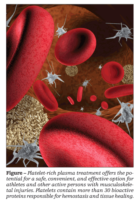
Platelets are formed in the bone marrow as nonnucleated cells, the end product of megakaryocytes.7 They contain several secretory granules (α, δ, λ), which contain more than 30 bioactive proteins and are responsible for hemostasis and tissue healing (Figure). The α-granules contain the majority of growth factors.
Once formed, platelets circulate in a resting state and have a life span of 7 to 9 days.8 Platelets secrete inflammatory and mitogenic proteins within 10 minutes of clot formation. Most of the growth factors are released within 1 hour of arrival at the injury site, although small amounts continue to be released over the following 7 days.2,9,10
Wound healing cascade
Signaling proteins at the injury site initiate the coagulation cascade with the development of the fibrin plug, platelet aggregation, and clot formation. Extracellular calcium stimulates degranulation of the platelet's α-granules in a process called “activation.”7 The activated platelets release growth factors, which direct chemotaxis of neutrophils, monocytes, and fibroblasts to the wound. Over the following few days, these growth factors and cell lines are thought to promote removal of tissue debris, angiogenesis, and the laying down of extracellular matrix.6
Formulation and delivery
PRP is derived from autologous anticoagulated blood. The anticoagulant prevents platelet activation from clot formation before the PRP is ready for therapeutic use; clot formation is necessary for the release of growth factors. PRP may be prepared in the clinic or operating department immediately before treatment.
Several methods are used to prepare PRP, including tabletop centrifugation, selective filtration, and plateletpheresis.6 PRP is stable in the anticoagulated form for up to 8 hours.9 To activate the PRP, 10% calcium chloride or thrombin is added to release the growth factors.8
Once the PRP is prepared, a dual lumen syringe may be used for simultaneous injection, via standard injection technique, of the 10:1 solution of PRP and calcium into the injury site.6 Some authors recommend ultrasonography-guided injections to ensure delivery of the aliquot to the precise site of injury.3
CURRENT CLINICAL
APPLICATIONS
PRP therapy currently is thought to be experimental and is not reimbursed by third-party payers.4 Data from several animal research models support its effectiveness, but extrapolating that data to safety and efficacy in human use is difficult. The theoretical and anecdotal benefits of growth hormone use have led to many human studies of treatment for common musculoskeletal injuries.
Lateral epicondylitis
Patients who have chronic severe lateral epicondylitis and for whom standard nonoperative treatment is not successful may consider surgery as the next therapeutic option. Mishra and Pavelko11 reported a small, prospective, nonrandomized, controlled study of 20 patients with chronic lateral epicondylitis who were treated with PRP instead of surgery. Eight weeks after therapy, 60% of PRP-treated patients reported improvement in symptoms compared with 16% of controls. A double-blind, prospective, randomized trial of 230 patents is being conducted.1
Peerbooms and associates12 conducted a randomized controlled study of treatment for lateral epicondylitis in 100 patients in 2 cohorts (PRP and local corticosteroid injection). Using the Visual Analog Scale (VAS) and the Disabilities of the Arm, Shoulder, and Hand (DASH) Outcome Measure, the study showed a greater improvement in both scores for the PRP group. The VAS scores showed a 49% improvement in the corticosteroid group compared with a 73% improvement in the PRP group. Similarly, the corticosteroid group DASH scores improved by 51% compared with a 73% improvement in the PRP group.
At 2-year follow-up, both groups showed improvement in their VAS scores, but the corticosteroid group DASH scores returned to baseline.13 DASH scores in the PRP group showed significant improvement.
Achilles tendinopathy
Achilles tendon rupture is a common problem among athletes. Healing is slow because of limited blood supply to the area, and surgery often is required.
de Vos and associates14 conducted a randomized controlled trial of PRP injection treatment in patients with chronic Achilles tendinopathy. The study group treated with PRP injection and exercise showed no greater improvement in symptoms than the control group treated with a saline injection and exercise. In a double-blind placebo-controlled study of treatment for chronic Achilles tendinopathy in 54 patients, de Jonge and colleagues15 found no significant difference in improvement between the cohort treated with concentric exercise and PRP injection and the control group treated with exercise and saline injection.
Snchez and colleagues16 reported a case-control study of 12 athletes who underwent surgical repair of Achilles tendon tears with intraoperative use of PRP. The athletes treated with PRP regained range of motion and returned to training activities earlier than the control group who did not receive the PRP during surgery.
Rotator cuff tears
Randelli and colleagues17 used PRP intraoperatively during arthroscopic rotator cuff repair in a small pilot study of 14 patients that evaluated its potential for repair of these injuries. There were no adverse effects, and patients' pain and functional scores improved postoperatively at 6, 12, and 24 months' follow-up.
Castricini and coworkers18 reported a recent randomized controlled study of 88 patients with small or medium-size rotator cuff tears. Patients were assigned to either of two cohorts: patients in one cohort underwent double row arthroscopic repair of the rotator cuff tear with PRP and patients in the other did not receive the augmentation. The authors observed no statistical difference in MRI tendon scoring between the two groups after treatment.
Patellar tendinosis
Chronic patellar tendinosis, or jumper knee, is a common overuse injury in athletes caused by repetitive loading of the patella tendon, resulting in microscopic tears. In a small prospective pilot study of 20 athletes with chronic jumper knee, Kon and coworkers19 administered a series of 3 intratendinous and peritendinous injections of PRP at 15-day intervals. The injections were made with multiple penetrations to the site of pain. At the 6-month follow-up, 6 athletes showed complete functional recovery, 8 showed marked recovery, 2 showed mild recovery, and 4 had no improvement in symptoms. The authors concluded that their results are encouraging for the use of PRP injections in the management of jumper knee, at least in the short term.
Plantar fasciitis
Chronic plantar fasciitis, a common cause of foot pain, involves tissue degeneration at the origin of the plantar fascia at the medial tuberosity of the calcaneus.20 Barrett and Erredge21 conducted a study of chronic refractory plantar fasciitis in which 9 patients were treated with ultrasonography-guided PRP injections into the medial plantar fascia. At 1 year, 77.9% of patients reported complete resolution of symptoms.
A randomized, controlled, multicenter trial is being conducted by Peerbooms and associates.22 The investigators have enrolled 120 patients with chronic plantar fasciitis for whom conservative treatment was not successful. They plan to compare corticosteroid injection with PRP injection.
Anterior cruciate
ligament (ACL) tears
Healing after operative reconstruction for ACL rupture may take many months. Radice and colleagues23 conducted a prospective single-blind study of 50 patients undergoing ACL reconstruction surgery in which one group had PRP added to the graft intraoperatively and the control group did not. The study results, based on MRI assessment, indicated a 48% shortening of time to complete homogeneous graft appearance with the use of PRP.
Degenerative cartilage
lesions of the knee
Treatment for degenerative changes in the knee has included oral NSAIDs, glucosamine and chondroitin sulfate, and intra-articular injections with hyaluronic acid or corticosteroids. Response to treatment with these modalities is varied and often temporary.
Kon and associates24 conducted a pilot study to evaluate the safety and efficacy of PRP treatment for degenerative lesions of the knee; 100 nonrandomized patients with chronic degenerative knee conditions received 3 intra-articular injections of PRP. The authors reported a statistically significant improvement of all clinical scores at 6 to 12 months' follow-up.
GROWTH HORMONE USE
IN ATHLETES
The World Anti-Doping Agency has addressed the use of growth factors, including IGF-1, a component of PRP preparations, and prohibits the use of intramuscular injection of autologous blood products that contain growth factors in elite athletes.2 The use of PRP delivered by other routes requires a Therapeutic Use Exemption (TUE) in compliance with international standards.3 However, the bureaucratic process involved with receiving a TUE significantly limits its application. The US Anti-Doping Agency also considers a PRP injection to be equivalent to an injection of growth factors.3
An International Olympic Committee 2008 consensus statement recommended further research into biologic therapies to ensure that they are optimized and safe for patients and for athletes.25
Professional sports in the United States do not fall under the jurisdiction of anti-doping agencies but are governed by league and union contracts. Several performance-enhancing substances have been banned, but PRP is not one of them.3
CONTRAINDICATIONS
AND COMPLICATIONS
Contraindications to the use of PRP include preexisting coagulopathies, concurrent anticoagulant therapy, active infection, tumor, metastatic disease, pregnancy, and hypersensitivity to bovine thrombin.4 Several systemic complications may be associated with the use of PRP. There is a theoretical concern that the injection of PRP could initiate a systemic increase in growth factors, resulting in a cancer-like effect; however, Foster and associates3 did not find any data to support this concern.
Infection is a potential complication of any invasive procedure. However, when the injection is performed with a sterile procedure, the autologous nature of the PRP minimizes the risk of transmissible infection or allergic reactions.
Healing of soft tissue injuries induces fibrosis or scar formation. Creaney and Hamilton2 raised the concern that the injection of growth factors may increase the risk of fibrosis, preventing full recovery. However, Snchez and colleagues26 maintained that growth factors are antifibrotic agents and may actually help reduce scar formation. Further study of this potential complication is needed.
DISCUSSION
The use of PRP to manage musculoskeletal injuries is gaining favor with orthopedists. A review of the early literature indicates that PRP has the potential to be effective for select tendon, ligament, and cartilage disorders. Concerns regarding safety are few, and researchers contend that they may be unfounded because the use of autologous blood obviates many of them.
The research data on the clinical use of PRP therapy are just beginning to be accumulated and involve several limitations. Published data on PRP applications for sports medicine injuries are limited by small study size, making discrimination between real and chance effects difficult.27 A variety of PRP studies have been conducted-some randomized controlled trials, some case-control studies, and some pilot studies-all using different formulations, treatment protocols, and posttreatment management. With the data that are available, generalizing the efficacy of PRP treatment is difficult, as is concluding for which conditions and under which clinical circumstances PRP is useful or contraindicated.
In addition, because the optimal concentration, dose range, and timing of therapy after injury have yet to be defined, comparing results from one study to the next is difficult. Defining the clinical effect for each type of tissue and determining whether PRP is equally effective in managing acute and chronic disease needs further study.
CONCLUSION
PRP treatment offers the potential for a safe, convenient, and effective therapeutic option for athletes and other active persons who have musculoskeletal injuries. Although study results are not conclusive, they have indicated generally positive results and a good safety profile with PRP treatment.
A wide range of clinical studies are being conducted, and investigators are expanding the study of PRP therapy to its effectiveness in other musculoskeletal disorders. Before clinicians can offer their patients this treatment with confidence, further clinical investigation focused on standardization of concentration, protocols for clinical applications in various settings, and the proper role of PRP treatment for sports medicine injuries in amateur and professional sports is needed.
References
1. Mishra A, Woodall J Jr, Vieira A. Treatment of tendon and muscle using platelet-rich plasma. Clin Sports Med . 2009;28:113-125.
2. Creaney L, Hamilton B. Growth factor delivery methods in the management of sports injuries: the state of play. Br J Sports Med. 2008;42:314-320.
3. Foster TE, Puskas BL, Mandelbaum BR, et al. Platelet-rich plasma: from basic science to clinical applications. Am J Sports Med. 2009;37:2259-2272.
4. Hall MP, Band PA, Meislin RJ, et al. Platelet-rich plasma: current concepts and application in sports medicine [published correction appears in J Am Acad Orthop Surg. 2010;18:17A]. J Am Acad Orthop Surg. 2009;17:602-608.
5. Lopez-Vidriero E, Goulding KA, Simon DA, et al. The use of platelet-rich plasma in arthroscopy and sports medicine: optimizing the healing environment. Arthroscopy. 2010;26:269-278.
6. Alsousou J, Thompson M, Hulley P, et al. The biology of platelet-rich plasma and its application in trauma and orthopaedic surgery: a review of the literature. J Bone Joint Surg. 2009;91B:987-996.
7. Rozman P, Bolta Z. Use of platelet growth factors in treating wounds and soft-tissue injuries. Acta Dermatovenerol Alp Panonica Adriat. 2007;16:156-165.
8. Mehta S, Watson TJ. Platelet rich concentrate: basic science and current clinical applications. J Orthop Trauma. 2008;22:432-438.
9. Marx RE. Platelet-rich plasma: evidence to support its use. J Oral Maxillofac Surg. 2004;62:489-496.
10. Kevy S, Jacobson M. Preparation of growth factor enriched autologous platelet gel. In: Proceedings from the Society of Biomaterials 27th Annual Meeting; April 2001; St Paul.
11. Mishra A, Pavelko T. Treatment of chronic elbow tendinosis with buffered platelet-rich plasma. Am J Sports Med. 2006;34:1774-1778.
12. Peerbooms JC, Sluimer J, Bruijn DJ, Gosens T. Positive effect of an autologous platelet concentrate in lateral epicondylitis in a double-blind randomized controlled trial: platelet-rich plasma versus corticosteroid injection with a 1-year follow-up. Am J Sports Med. 2010;38:255-262.
13. Gosens T, Peerbooms JC, van Laar W, den Oudsten BL. Ongoing positive effect of platelet-rich plasma versus corticosteroid injection in lateral epicondylitis: a double-blind randomized controlled trial with 2-year follow-up. Am J Sports Med. 2011 Mar 21; [Epub ahead of print].
14. de Vos RJ, Weir A, van Schie HT, et al. Platelet-rich plasma injection for chronic Achilles tendinopathy: a randomized controlled trial. JAMA. 2010;303:144-149.
15. de Jonge S, de Vos RJ, Weir A, et al. Platelet-rich plasma for chronic Achilles tendinopathy: a double-blind randomised controlled trial with one year follow-up. Br J Sports Med. 2011;45:e1. doi:10.1136/bjsm.2010.081554.40.
16. Snchez M, Anitua E, Azofra J, et al. Comparison of surgically repaired Achilles tendon tears using platelet-rich fibrin matrices. Am J Sports Med. 2007;35:245-251.
17. Randelli PS, Arrigoni P, Cabitza P, et al. Autologous platelet rich plasma for arthroscopic rotator cuff repair: a pilot study. Disabil Rehabil. 2008;30:1584-1589.
18. Castricini R, Longo UG, De Benedetto M, et al. Platelet-rich plasma augmentation for arthroscopic rotator cuff repair: a randomized controlled trial. Am J Sports Med. 2011;39:258-265.
19. Kon E, Filardo G, Delcogliano M, et al. Platelet-rich plasma: new clinical application: a pilot study for the treatment of jumper's knee. Injury. 2009;40:598-603.
20. Buchbinder R. Clinical practice: plantar fasciitis. N Engl J Med. 2004;350:2159-2166.
21. Barrett SL, Erredge SE. Growth factors for chronic plantar fasciitis? Podiatry Today. 2004;17(11):37-42.
22. Peerbooms JC, van Laar W, Faber F, et al. Use of platelet rich plasma to treat plantar fasciitis: design of a multi centre randomized controlled trial. BMC Musculoskelet Disord. 2010;11:69.
23. Radice F, Ynez R, Gutirrez V, et al. Comparison of magnetic resonance imaging findings in anterior cruciate ligament grafts with and without autologous platelet-derived growth factors. Arthroscopy. 2010;26:50-57.
24. Kon E, Buda R, Filardo G, et al. Platelet-rich plasma: intra-articular knee injections produced favorable results on degenerative cartilage lesions. Knee Surg Sports Traumatol Arthrosc. 2010;18:472-479.
25. Ljungqvist A, Schwellnus MP, Bachl N, et al. International Olympic Committee consensus statement: molecular basis of connective tissue and muscle injuries in sport. C lin Sports Med. 2008;27:231-239, x-xi.
26. Snchez M, Anitua E, Orive G, et al. Platelet-rich therapies in the treatment of orthopaedic sport injuries. Sports Med. 2009;39:345-354.
27. Hackshaw A. Small studies: strengths and limitations. Eur Respir J. 2008;32:1141-1143.

Real-World Study Confirms Similar Efficacy of Guselkumab and IL-17i for PsA


