Publication
Article
Family Practice Recertification
Newborn with Facial Marks
Author(s):
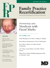
This newborn was noted to have these facial lesions at time of birth. On further exam there is some erythema on the nape of the neck. The parents indicate that the color seems to be more or less prominent at various times. The pregnancy and delivery were otherwise uncomplicated.
This newborn was noted to have these facial lesions at time of birth. On further exam there is some erythema on the nape of the neck. The parents indicate that the color seems to be more or less prominent at various times. The pregnancy and delivery were otherwise uncomplicated.
What is your diagnosis?
A. Nevus simplex (Angel kisses)
B. Nevus flammeus (Port wine stain)
C. Strawberry hemangioma
D. Cherry hemangioma
This infant’s lip lesion is fairly dense in color similar to nevus flammeus, but the pattern of several lesions which are bilateral, affecting the eyelids and also on the nape of the neck are classic for nevus simplex.
Therefore and fortunately this child has “Angel kisses” and not a port wine stain. This very common finding is also sometimes called salmon patches or capillary malformation although it is debated if it is a true malformation or simply a dilation of capillaries. The natural course of nevus simplex is benign with the facial lesions lessening in prominence over the first year. The lesions on the nape of the neck often called “stork bites” tend to be permanent, but due to the location are usually only noticeable with very short hair or hair loss1.
Nevus Flammeus with the common name “port wine stain” denotes two clinically important features: First the deep red color of red wine from Portugal and secondly the stain reference due to its permanent nature. It typically presents unilaterally along the nerve distribution patterns of the face. The color is quite prominent and does not spontaneously resolve. Over time the lesions can even become nodular.
Involvement above the lateral canthus of the eye can be associated with Sturge Weber syndrome including glaucoma and intracranial abnormalities. If there are no underlying abnormalities no treatment is required, but the large deep red lesions can be very prominent and disconcerting for the patient. They can be covered with thick opaque make up or pulse dye laser in the first year of life achieves good to excellent results in half of treated patients2.
Strawberry hemangiomas are usually not visible at birth or only faintly present. Over the first year of life they subsequently grow into raised lesions often with an irregular surface looking like a strawberry. They spontaneously involute over the next 6-7 years with partial or complete resolution. Large lesions, eroding lesions or those affecting sensitive areas around the eyes or mouth are now treated with oral propranolol with improvement in 98% of lesions3.
Cherry angiomas are not present at birth but develop in adulthood as bright red macules with some enlarging to raised lesions several millimeters in diameter. They are benign but can be treated with electrosurgery, cryosurgery or pulsed dye laser for cosmetic purposes.
About the authors:
Daniel Stulberg, MD, is a Professor of Family and Community Medicine at the University of New Mexico. After completing his training at the University of Michigan, he worked in private practice in rural Arizona before moving into full-time teaching. Stulberg has published multiple articles and presented at many national conferences regarding skin care and treatment. He continues to practice the full spectrum of family medicine with an emphasis on dermatology and procedures.


