Publication
Article
Internal Medicine World Report
Ultrasound Guides Tailored Treatment for Common Shoulder Disorders
Author(s):
Although the true prevalence of shoulder disorders in general practice remains unknown, an analysis of ultrasound imaging reports offers physicians some insight into the underlying causes of shoulder pain.

Although the true prevalence of shoulder disorders in general practice remains unknown, an analysis of ultrasound imaging reports published in BMC Family Practice offers physicians some insight into the underlying causes of shoulder pain.
“As ultrasound is an easily accessible, accurate, relatively cheap, and non-invasive and non-ionising imaging procedure, general practitioners (GPs) frequently refer patients for ultrasound, resulting in a rise in test volume of 100% over the last 6 years,” the study authors noted. “However, at present, there is no clear evidence for using ultrasound for acute shoulder pain.”
With a primary objective to “determine the frequency of the specific ultrasound-diagnosed shoulder disorders of patients with shoulder pain referred from general practice,” Ramon P.G. Ottenheijm, GP/PhD, MD, and colleagues from the Netherlands retrospectively reviewed 240 ultrasound shoulder examinations performed at a hospital in 2011 based on referrals by GPs.
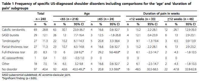
While no shoulder disorders were found on ultrasound for the majority of patients aged <65 years, significantly more full-thickness tears were seen in those aged ≥65 years, the researchers reported. Across the entire patient population, ultrasound imaging most frequently displayed “calcific tendonitis (29%), followed by subacromial-subdeltoid (SASD) bursitis (12%), tendinopathy (11%), partial-thickness tears (11%), full-thickness tears (8%), and acromioclavicular (AC) osteoarthritis (0.4%),” the authors wrote (Table 1).

In a subgroup of 41 patients with 2 disorders diagnosed via ultrasound, calcific tendonitis was again the most prevalent condition, and it was most frequently seen in combination with tendinopathy or SASD bursitis. In fact, 43% of patients with SASD bursitis “had concomitant calcific tendonitis, with a specific calcific bursitis in 10%,” the investigators noted.
“Contrary to findings from secondary care, calcific tendonitis is the most common ultrasound-diagnosed disorder affecting patients who are symptomatic enough and have had symptoms for a sufficient duration to warrant referral by their GP for ultrasound,” the authors concluded, asserting their new findings “imply that patients can be stratified into diagnostic subgroups, allowing more tailored treatment than currently applied” in daily practice.


